|
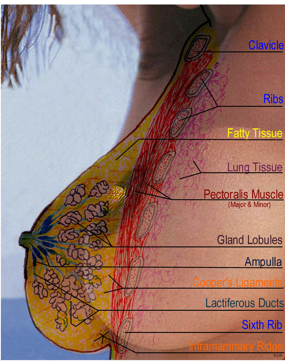 Clavicle Bone
The clavicle is the prominent bone that goes across your upper chest,
from shoulder to shoulder, above the breasts and below the neck. We often
refer to it as our collar bone. It is the upper delimiter of the breast.
It has a distinctive notch in the center, directly above the sternum and
below the trachea.
Ribs
When breast implants are used, they may either be located between the
Pectoral muscles and the ribs (submuscular), or in front of the Pectoral muscles, but
behind the breast tissues (subglandular). The lower attachment of the breast to the chest wall
is usually near the sixth rib when a breast reaches full maturity at the
end of puberty. This attachment point (in relation to the
sixth rib) actually changes over a woman's lifetime, moving in the downward
direction as she ages. Note that this is only the attachment point, and
the breast ptosis or sagging is a totally separate issue.
Fatty Tissue
One third of the average sized adult breast of a non-lactating woman consists
of fatty tissue. Larger adult breasts usually only contain more fatty
tissue. Smaller than average breasts still contain the same amount of
Glandular (milk producing) tissue, but have less fatty tissue. Poor nutrition
will prevent the storage of excess fat in the breasts, and may result
in a sagging (pendulous) effect.
Pectoralis (Pectoral) Muscles
These connect between the chest and the arms. They may be affected or
partially removed during a radical mastectomy, affecting the use of the
arm that is involved. These muscles are what will develop when chest exercises
are done to increase breast size. A minimal effect to the breast size
will result, with a possibility of a negative overall effect due to an
increase in physical activity during the special exercises removing some
of the fatty tissues from the breasts.
Gland Lobules
Also known as acini or alveoli, these are the actual source of breast
milk. They develop during puberty to a certain degree, and then complete
their development during pregnancy. Lobular development causes the breasts
to expand. The final development occurs during pregnancy, and allows the
production of milk after giving birth. Most breasts of the same cup size have the same
average number
of lobules, and they are grouped into 15 to 25 lobes. As a woman approaches
menopause, the lobules start to atrophy, allowing a reduction in the density
of the breasts. They may be replaced with fatty tissue, further softening
the breast; a process called involution.
Ampulla
(Lactiferous Sinus)
This is an enlarged area in the Lactiferous Duct that maintains a small
reserve of milk that will be immediately available to the infant that
is placed at the breast. It will maintain his/her interest until the Lobules
are able to produce and release more milk. Pressure is placed directly
onto these to manually express milk from the breast, not on the nipple.
Suspensory Ligaments
Often referred to as Cooper's Ligaments, these are the fibrous
connections between the inner side of the breast skin and the pectoral
muscles. Working in conjunction with the fatty tissues and the more fibrous
lobular tissues, they are largely responsible for maintaining the shape
and configuration of the breast. They bear a major portion of the task
of preventing breast ptosis (sagging).
Lactiferous Ducts (milk ducts)
These gather the milk from the lobules and deliver it to the nipple. Each
lobe usually has its own ductal system.
Inframammary Ridge
Located at the junction between the lower side of the breast and the chest
wall, it is a more dense area that is easily felt when lying on your
back. This will often thicken with age. It might be (and often is) confused with a mass or lump during a
Breast Self-Examination.
 |