|
|
|

|
Lymphatic System
|
"You mean there is more than blood and milk circulating in my breasts?" |
|
|
|
|
Lymphatic System
|
"You mean there is more than blood and milk circulating in my breasts?" |
|
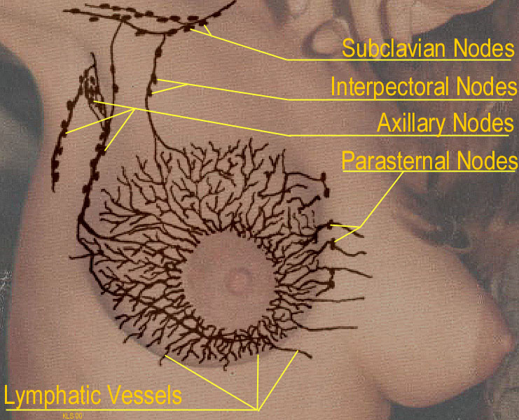
Lymph Nodes or Glands Currently, a process called Sentinel Node Biopsy (SNB) is being used to reduce the number of lymph nodes that have to be removed during a lymph node biopsy. It is not that simple to remove a specific number of lymph nodes, due to the difficulty of finding them hidden among the fatty tissues. In a SNB, a dye is injected directly into the location of the suspicious mass in the breast, and the lymphatic vessels that are charged with the duty of cleansing that particular location will pick up that dye after a short while and take it back to their node. When the biopsy is done, the node with the dye is more likely to be identifiable, making the removal of a larger number of nodes unnecessary. The thought behind this is that when a malignancy in the breast (or any other part of the body for that matter) starts to metastasize (send off "seeds" or loose cells to start cancers elsewhere in the body) the lymphatic system will sweep them up and take them to the associated lymph node. The same thing happened to the dye that was injected. Once the associated lymph node is found, it is excised (removed surgically) and dissected under a microscope to see if it has any of those wandering malignant cells. If ANY cells have left the malignant mass, it is extremely likely that the associated lymph node will have picked some of them up. if one or more malignant cells are found, more have likely been released and chemo-therapy most likely will be prescribed. if no malignant cells are found in the "sentinel node", it is accepted that there have most likely not been any malignant cells released from the breast mass, and chemo-therapy will probably NOT be prescribed. If you are facing a lymph node dissection, I strongly suggest that you ask your surgeon about having a Sentinel Node Biopsy. The benefits of this are a much lower likelihood of developing lymphedema in the associated arm. When lymphatic fluids are not properly circulated, due to restriction from clothing or the use of a bra, the lymphatic fluid enters the condition referred to as "Stasis" where it literally becomes static. Any breast cells that through a "genetic accident" becomes malignant needs to be carried away and disposed of. That is what the lymphatic system is designed to do. If a static or stasis condition exists, that malignant breast cell can go ahead and start developing right where it lies, and that is where breast cancer starts. You can read more about it in several articles located on the Suspected Causes Overview".
|
 Lymphatic Vessels